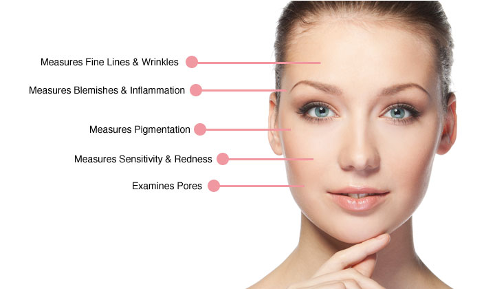
If you are curious about what lies beneath the surface of your skin, a detailed skin analysis will reveal anomalies that are not visible to the naked eye.
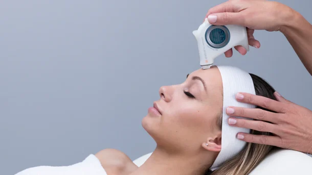
Everyone’s skin is different. So how do we accurately diagnose a person’s skin type and skin problems so that we can treat them? This is where an advanced diagnostic tool, the Skin Analyzer, comes in handy to provide you with a skin analysis!
As a patient, if you know your skin, you can better understand the reasons for the skincare products or treatments recommended to you. Your skin is digitally recorded before and after treatment so you can see for yourself if your skin has improved.
How do I prepare my skin for a skin analysis?
The skin needs to be in its natural state, free of any products, so that it can be analyzed accurately. Therefore, all you need to do is cleanse it with the appropriate cleanser and not apply anything to it for 1-2 hours. This ensures that the skin is restored to its natural state of oil levels, pores, bacteria levels, etc. so that it can be analyzed.
What do the images show?
A daylight image is a “control” image. That is, it was taken without interference from indoor daylight or artificial light. This is the “real” image that we use to compare with other images generated by the analyzer.
The cross-polarized image probes for blood vessels and pigmentation under the surface. This will show any skin inflammation and/or broken capillaries.
A parallel polarized image enhances the texture of the skin surface. In this image, textural irregularities, fine lines and wrinkles are clearly visible. It also identifies over-hardened skin, blackheads and enlarged pores.
True UV Imaging uses harmless UV light to penetrate the surface of the skin, thus turning human skin into a light. It produces very clear images compared to other handheld devices. Skin problems exposed include melasma, sun damage, freckles, thinning skin, dry skin, clogged pores, normal pores and normal oil flow. Since porphyrins produced by bacteria fluoresce in this light, normal bacteria on the surface of the skin are highlighted by bright red spots.
Wood’s light images are specifically designed to look at the activity of the sebaceous glands. This can reveal oily, dry or combination skin.
The skin color image is a combination of all the other images. It can show changes in skin color in different areas of the face due to sun damage.
Why is this analysis important?
Skin analysis is important because images can be used to differentiate between diagnoses that are difficult to distinguish. For example, melasma, sun damage and post-inflammatory hyperpigmentation can be difficult to distinguish with the naked eye.
It is ingenious to be able to diagnose surface secretions, surface texture, inflammation and skin infections in less than a minute of analysis.